Spatially encoded biological assays
Patent No. US10983113 (titled "Spatially encoded biological assays") on Apr 1, 2020. The application was issued on Apr 20, 2021.
What is this patent about?
’113 is related to the field of spatially encoded biological assays and high-resolution molecular mapping. In traditional genomics and proteomics, the spatial context of biological molecules is often lost during sample homogenization, making it difficult to understand how gene expression or protein distribution varies across different cell types within a complex tissue. The invention addresses the need for a scalable, multiplexed system that can simultaneously identify the presence, abundance, and precise location of numerous biological targets within a solid sample.
The underlying idea behind ’113 is the decoupling of target detection from spatial localization through a programmable encoding scheme. Instead of relying on physical separation or manual microdissection, the invention utilizes probes that bind to specific biological targets and are subsequently linked to unique coding tags. These tags act as digital coordinates, allowing researchers to pool assay products for high-throughput analysis while retaining the ability to map every data point back to its original position in the tissue architecture.
The claims of ’113 focus on a method for determining the presence or abundance of a target protein at a specific region of interest within a tissue sample. This process involves delivering a plurality of probes—each consisting of a capture agent like an antibody conjugated to an oligonucleotide—to the sample. A critical step in the claimed method is the use of imaging to identify the specific region of interest, followed by the removal and sequencing of the oligonucleotides from that targeted area to quantify the protein targets present.
In practice, the system works by applying these conjugated probes to a tissue section affixed to a support. After the capture agents bind to their respective protein targets, the sample is imaged to define the boundaries of specific histological features or cell populations. The oligonucleotides associated with the bound probes are then recovered from these defined regions. By determining the sequence of these oligonucleotides, the system generates a digital readout that correlates the frequency of specific sequences with the concentration of proteins at the imaged location.
This approach differs from prior methods by combining the high-multiplexing capabilities of next-generation sequencing with the visual precision of microscopy. Unlike laser capture microdissection, which is labor-intensive and difficult to scale, or standard in situ hybridization, which is limited in the number of targets it can detect simultaneously, this invention allows for the analysis of thousands of different proteins or transcripts in a single workflow. It effectively transforms a physical tissue sample into a searchable digital map of molecular activity.
How does this patent fit in bigger picture?
Technical Landscape
In the early 2010s when ’113 was filed, comprehensive gene expression and protein analysis were typically implemented using microarrays, serial analysis of gene expression (SAGE), or high-throughput qPCR. While these methods enabled quantitative analysis of many sequences per sample, systems commonly relied on bulk processing or laser capture microdissection rather than automated, high-resolution spatial mapping. At a time when hardware and software constraints made the simultaneous measurement of hundreds of thousands of targets across distinct spatial locations non-trivial, researchers were often forced to choose between high levels of multiplexing and the preservation of the native spatial context of the biological sample.
Prosecution Position
The disclosed invention represents a meaningful technical advancement through the integration of a spatial encoding scheme with high-throughput digital sequencing to enable high-resolution mapping of biological targets. The architectural shift involves delivering encoded probes to a sample in known spatial patterns, where each probe contains a coding tag that identifies the specific delivery site. This structural solution allows for the pooling and parallel sequencing of vast numbers of probes while maintaining the ability to computationally associate each target's abundance or activity back to its original location in the tissue. This capability overcomes the technical constraint of low spatial resolution inherent in physical transference methods, enabling the simultaneous analysis of millions of biological targets across a sample surface.
Claims
This patent contains a total of 30 claims, with claim 1 serving as the sole independent claim. The independent claim focuses on a method for determining the presence or abundance of target proteins within a specific region of interest in a tissue sample by utilizing capture agents conjugated to oligonucleotides, imaging the sample to identify the region, and subsequently removing and sequencing the oligonucleotides. The dependent claims serve to further define the technical parameters of the process, including specific imaging techniques like immunohistochemistry, methods for separating unbound probes, the use of fluorescent labels or hybridization for detection, the identification of specific subcellular targets, and the application of the method to multiple unique proteins or various tissue preparation types such as fresh-frozen or formalin-fixed paraffin-embedded sections.
Key Claim Terms New
Definitions of key terms used in the patent claims.
Litigation Cases New
US Latest litigation cases involving this patent.
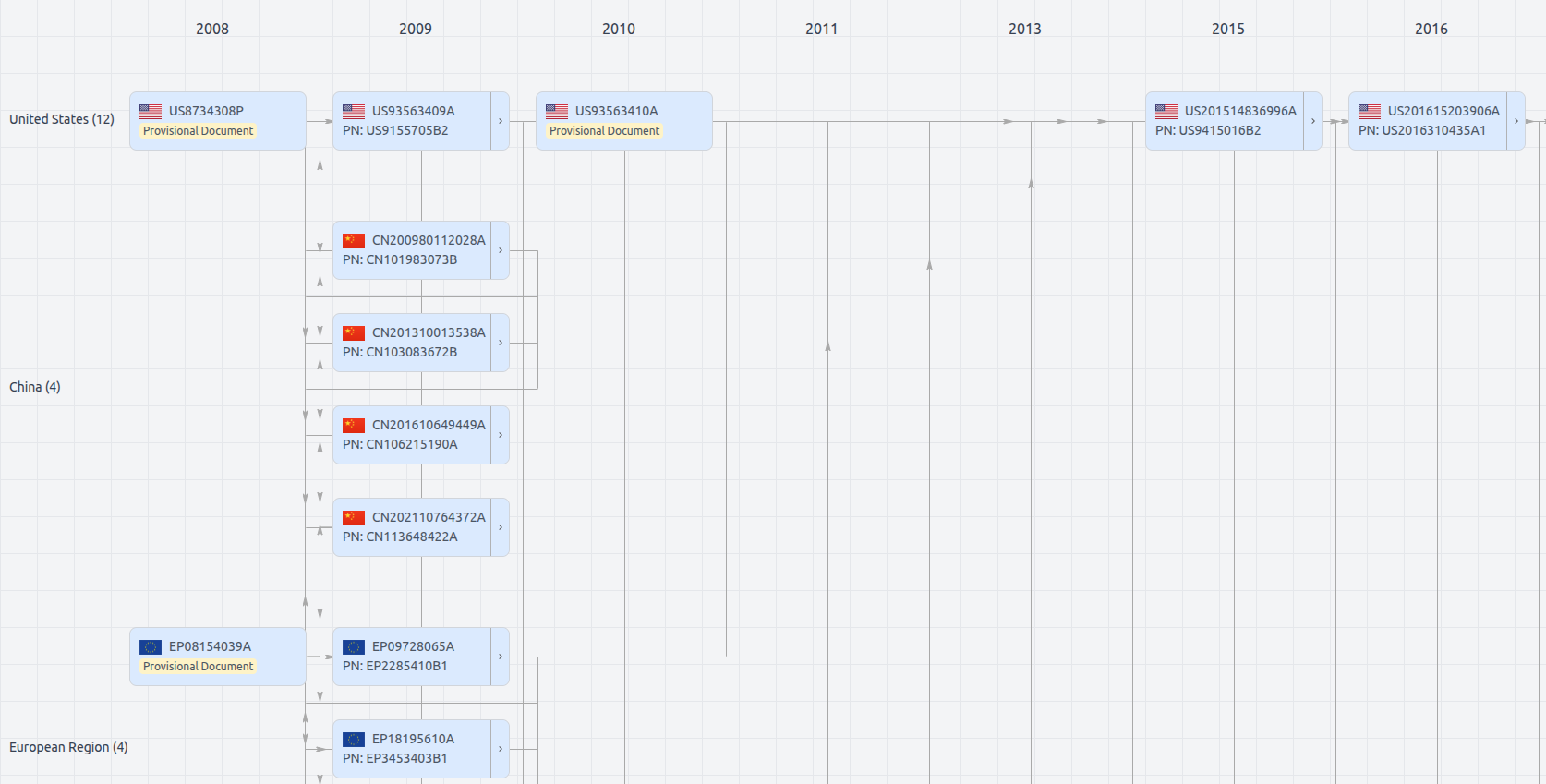
Patent Family
File Wrapper
The dossier documents provide a comprehensive record of the patent's prosecution history - including filings, correspondence, and decisions made by patent offices - and are crucial for understanding the patent's legal journey and any challenges it may have faced during examination.
US10983113
- Application Number
- US16837924A
- Filing Date
- Apr 1, 2020
- Publication Date
- Apr 20, 2021
- External Links
- Slate, USPTO , Google Patents