Determining biomarkers from histopathology slide images
Patent No. US10957041 (titled "Determining biomarkers from histopathology slide images") on Mar 25, 2020. The application was issued on Mar 23, 2021.
What is this patent about?
’041 is related to the field of digital pathology and automated biomarker detection. Specifically, it addresses the technical challenge of identifying and quantifying cancer-related biomarkers, such as PD-L1, tumor-infiltrating lymphocytes, and molecular subtypes, directly from standard hematoxylin and eosin (H&E) stained histopathology images. The invention seeks to replace or supplement time-consuming and resource-intensive immunohistochemistry (IHC) staining and molecular sequencing with efficient, deep-learning-based analysis of digitized tissue slides.
The underlying idea behind ’041 is the use of specialized deep learning architectures to extract molecular and cellular insights from morphological features that are often invisible to the human eye. The system leverages two primary strategies: a multiscale deep learning framework that integrates broad tissue-level context with high-resolution cell segmentation, and a multiple instance learning (MIL) approach that allows the model to learn from slide-level labels (like RNA sequencing data) without requiring exhaustive, pixel-by-pixel manual annotations by pathologists.
The claims of ’041 focus on a computer-implemented method that separates a digital H&E image into a plurality of tiles and applies them to a deep learning framework to predict biomarker presence. Independent claim 1 specifically protects a multiscale approach that combines tissue classification for each tile with a cell segmentation model to identify individual cells, using the intersection of these data points to predict biomarker status. Claims 23, 26, and 27 focus on a single-scale framework utilizing a trained MIL controller to selectively or randomly discard tiles based on inferred class status, effectively filtering the input data to focus the neural network on the most predictive regions of the tissue.
In practice, the invention functions by processing high-resolution whole-slide images through a pipeline that first normalizes the data to account for staining variations across different labs. For multiscale implementations, the system identifies cell boundaries and interiors using a three-class segmentation model, which allows for accurate cell counting even in dense clusters. For molecular biomarkers like Consensus Molecular Subtypes (CMS), the system uses RNA transcriptome counts to cluster training data, allowing the image-based model to learn the visual signatures associated with specific genetic profiles. This enables the generation of a digital overlay report that maps the probability of biomarker presence across the entire specimen.
This approach differs from prior methods by eliminating the need for dense, localized annotations and specialized IHC staining for every patient. By utilizing weakly supervised learning, the system can be trained on vast datasets where only the overall patient outcome or genetic status is known, rather than requiring a pathologist to manually outline every tumor region. Furthermore, the use of a multi-field of view strategy ensures that the model considers both the immediate cellular morphology and the surrounding stromal environment, leading to more robust predictions of how a patient might respond to specific immunotherapies.
How does this patent fit in bigger picture?
Technical Landscape
In the late 2010s when ’041 was filed, the digitization of histopathology slides into high-resolution whole slide images was becoming a standard practice, at a time when diagnostic assessment was typically implemented using manual visual inspection by pathologists to identify tumor characteristics and biomarkers. While deep learning applications were emerging in medical imaging, systems commonly relied on traditional convolutional neural networks that assigned a single class label to an entire input image rather than performing granular, multi-class segmentation across large-scale digital slides. Furthermore, the massive pixel density of whole slide images made the use of standard fully convolutional networks for pixel-level classification non-trivial due to extreme computational redundancy and prohibitive processing time constraints.
Prosecution Position
The disclosed invention represents a meaningful technical advancement through the integration of a multiscale deep learning framework that combines tile-level tissue classification with pixel-level cell segmentation to predict biomarker presence. This architectural shift addresses the technical problem of computational inefficiency in large-scale image analysis by utilizing a tiling process and selective inference to reduce redundant calculations while maintaining high-resolution accuracy. The system enables the automated detection and quantification of biomarkers—such as tumor-infiltrating lymphocytes and PD-L1 status—directly from hematoxylin and eosin (H&E) stained images, overcoming the technical constraint of requiring labor-intensive immunohistochemistry (IHC) staining or manual pixel-by-pixel annotation for diagnostic reporting.
Claims
This patent contains 30 claims, with claims 1, 23, 26, and 27 serving as the independent claims. The independent claims focus on computer-implemented methods for identifying biomarkers in digital images of stained tissue slides by partitioning images into tiles and applying them to deep learning frameworks, including multiscale and single-scale models, to predict biomarker presence and generate visual reports. The dependent claims serve to specify particular tiling techniques, define specific biomarker types such as TILs and PD-L1, detail the architectures of the neural networks and segmentation models, and outline the training processes involving molecular datasets and tile selection criteria.
Key Claim Terms New
Definitions of key terms used in the patent claims.
Litigation Cases New
US Latest litigation cases involving this patent.
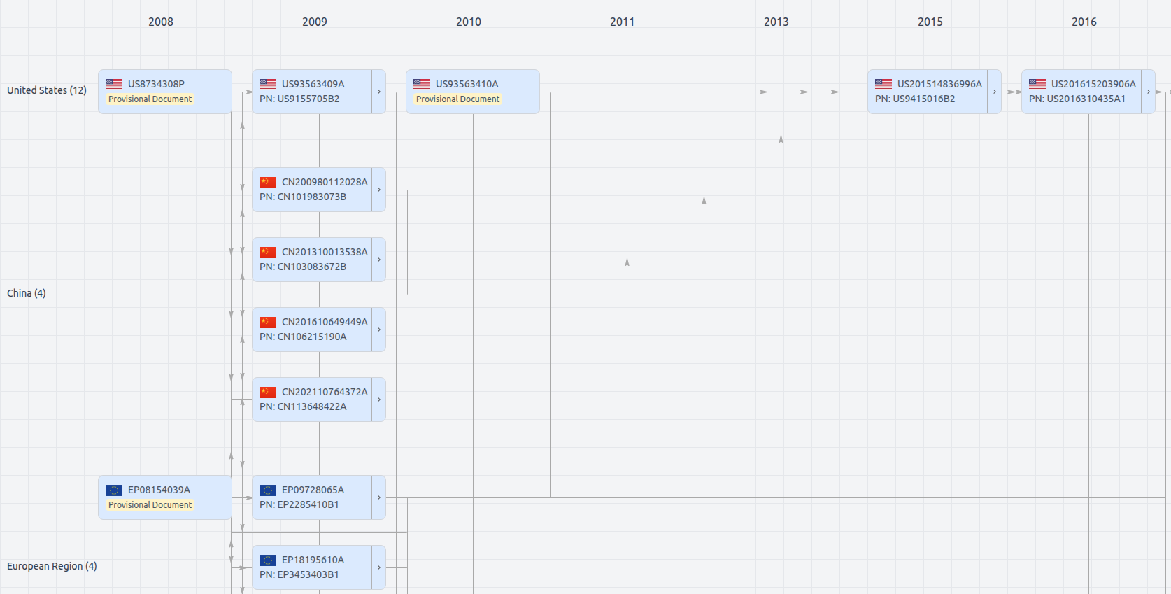
Patent Family
File Wrapper
The dossier documents provide a comprehensive record of the patent's prosecution history - including filings, correspondence, and decisions made by patent offices - and are crucial for understanding the patent's legal journey and any challenges it may have faced during examination.
US10957041
- Application Number
- US16830186A
- Filing Date
- Mar 25, 2020
- Publication Date
- Mar 23, 2021
- External Links
- Slate, USPTO , Google Patents