Artificial Intelligence Segmentation Of Tissue Images
Patent No. US10991097 (titled "Artificial Intelligence Segmentation Of Tissue Images") was filed by Tempus Ai Inc on Dec 31, 2019.
What is this patent about?
’097 is related to the field of medical image analysis, specifically the automated analysis of histological slides. The background involves the manual inspection of tumor samples by pathologists, a process that is time-consuming and subjective. Digital pathology, using high-resolution images, offers the potential for AI-driven analysis, but existing methods like Convolutional Neural Networks (CNNs) and Fully Convolutional Networks (FCNs) have limitations in handling the complexity and size of slide images, requiring excessive computation or pixel-level annotation.
The underlying idea behind ’097 is to efficiently classify tissue types in digital pathology slides by combining tile-based analysis with contextual information. Instead of processing each tile independently, the invention uses a multi-tile analysis that considers a larger region around each tile to incorporate structural tissue features. This approach reduces computational redundancy and improves classification accuracy by leveraging the spatial relationships between different tissue regions.
The claims of ’097 focus on methods for creating overlay maps and classifying tissue in digital images of slides. Claim 1 covers receiving a digital image, dividing it into tiles, and then, for each tile, identifying both local features within the tile and broader structural tissue features in a larger surrounding area. The tissue class for each tile is then determined based on both sets of features. Claim 6 builds on this by incorporating cell object detection and classification, overriding the tile classification with the cell classification when a cell object is present in a tile.
In practice, the invention segments a digital slide image into a grid of tiles. For each tile, a convolutional neural network (CNN) , specifically a tile-resolution fully convolutional network (FCN) like PhiNet, analyzes the tile itself and a larger surrounding region. This network is trained to recognize patterns indicative of different tissue classes (e.g., tumor, stroma, immune cells). The output is an overlay map where each tile is colored according to its predicted tissue class, providing a visual representation of the tissue composition.
The key differentiation from prior approaches lies in the multi-tile analysis and the use of a tile-resolution FCN. Traditional CNNs process images as a whole, while pixel-resolution FCNs require extensive pixel-level annotation. By analyzing tiles and their surrounding context, ’097 achieves a balance between computational efficiency and accuracy. Furthermore, the invention can incorporate cell-level information, refining the tissue classification by identifying and classifying individual cells within the tiles, leading to a more detailed and accurate analysis of the slide.
How does this patent fit in bigger picture?
Technical landscape at the time
In the late 2010s when ’097 was filed, digital pathology was gaining traction at a time when whole slide imaging was typically implemented using high-resolution scanners and analyzed using software tools that often relied on convolutional neural networks (CNNs) or fully convolutional networks (FCNs). However, when systems commonly relied on processing entire images or large sections at once, hardware or software constraints made pixel-level classification and analysis of large digital pathology images non-trivial due to the computational demands of processing such high-resolution data.
Novelty and Inventive Step
The examiner allowed the claims because the invention includes distinct features not found in the closest prior art (Tunstall). Specifically, Tunstall does not disclose "each tile of the plurality of tiles containing a respective portion of the digital image of the slide; and for each tile of the plurality of tiles: identifying features of the tile; identifying structural tissue features of a second portion of the digital image of the slide including at least part of one or more other tiles of the plurality of tiles, wherein the second portion is larger than the respective portion of the digital image contained in the tile; and identifying the majority class of tissue visible within the tile based at least in part on the features of the tile and the structural tissue features of the second portion of the digital image of the slide."
Claims
This patent contains 18 claims, with claims 1 and 6 being independent. The independent claims are directed to methods for creating overlay maps and tissue classification of digital images of slides, respectively, both involving tile-based analysis and identification of structural tissue features. The dependent claims generally elaborate on the independent claims by specifying further details, additional steps, or particular features related to the overlay generation, display, and cell object classification.
Key Claim Terms New
Definitions of key terms used in the patent claims.
Litigation Cases New
US Latest litigation cases involving this patent.
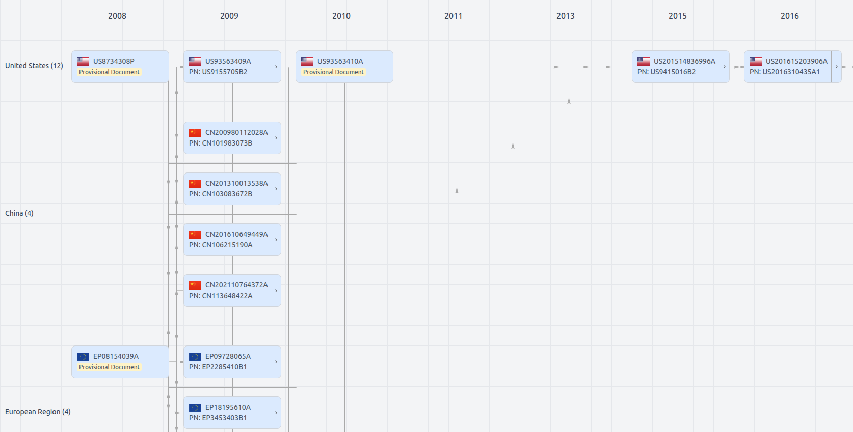
Patent Family
File Wrapper
The dossier documents provide a comprehensive record of the patent's prosecution history - including filings, correspondence, and decisions made by patent offices - and are crucial for understanding the patent's legal journey and any challenges it may have faced during examination.
Get instant alerts for new documents
US10991097
- Application Number
- US16732242
- Filing Date
- Dec 31, 2019
- Status
- Granted
- Expiry Date
- Dec 31, 2039
- External Links
- Slate, USPTO , Google Patents